Our Solution
Our Device Will...
Provide a high resolution picture of the surface of the mole
Give doctors key information about biological makeup of the mole
Provide a 3D image of the mole under the skin
Our Technology
Our technology is based on using spatially modulated illumination to characterize tissue optical properties of pigmented lesions/mole. We are using a specific structured illumination technique to gather information about absorption and scattering of the mole. Meaning we are able to obtain specific information about the mole using only red, blue, and green that will be beneficial to Dermatologists.
Our device will be able to receive and analyze the scattering and absorbance levels of a mole. The absorbance information can give information about the levels of Melanin and Hemoglobin in the mole. The scattering information can give information about the structure of the mole, specifically information about the collagen. Using this special illumination technique will also allow for analyzing the depth of the mole.
Benefits Of Our Device
Based on our technology our device will add additional features that Dermatologists can use to evaluate moles compared to the standard dermatoscope. The structured illumination technique will give doctors key information about the biological makeup of the mole. The levels of Melanin and Hemoglobin have been shown in literature that they can be used as indicators of skin cancer. Along with the structure of collagen, having abnormal structuring of collagen can also be an indicator of cancer. Our device will also allow Dermatologists to see a 3D image of what the mole looks like beneath the skin. Knowing the depth and structure of the mole can also be used to evaluate mole for potential skin cancer. We believe that a combination of all these benefits will allow doctors to better understand the moles they are evaluating thus reducing the number of unneeded biopsies.
Our Current Prototype and Results
We currently have a working bench top prototype that we are able to use to collect data and get preliminary results. The prototype is comprised of an outer shell that houses the main components of our device. The outer shell of our device is shown on the left and the main components are shown on the right.


Preliminary results show that we can get results about the absorbance and scattering of a mole as well as getting information about the depth of the mole. See on the left is an example of the scattering of the mole and on the right is an example of the preliminary depth information for 3D rendering of the mole.


Future work is required to improve results and validate these results.
Future Work
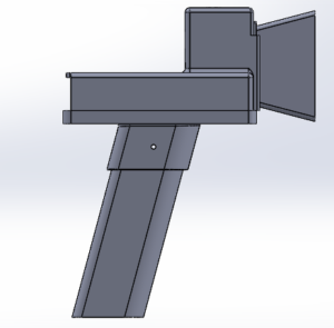
Future work includes improving the design of our device. A proposed design is shown below of how the outer shell could be change to make our device more ergonomic.


Future work will also include combining our main components to make our device smaller and more efficient. We will also we working to improve our data analysis to get better results regarding the levels of Melanin, Hemoglobin, structure of the Collagen, and work on developing our 3D view of the mole.
**This is currently an investigational device that is not yet for sale. Regulatory approval will be needed before this device becomes commercially available.

